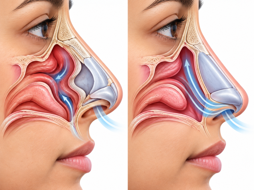
Rhinophyma is a thickening of the nose skin and oil glands that develops in some people with long-standing rosacea. It can make the nose appear larger, redder and more uneven. Medicines may help control early inflammatory changes, but established rhinophyma often needs a procedure to remove excess tissue and reshape the nose. A surgeon may use a scalpel, CO₂ laser or electrosurgery, depending on the severity and clinical findings.
What is rhinophyma and who gets it?
Rhinophyma is a form of phymatous rosacea. The skin around the nose gradually becomes thicker, and the oil glands and surrounding tissue may enlarge. The surface can develop prominent pores, an uneven texture and rounded or lobulated bumps.
The changes usually develop gradually. In more advanced cases, excess tissue can alter the shape of the nasal tip and sides. Severe enlargement may also interfere with airflow through the nostrils.
Rosacea can cause flushing, persistent redness, visible blood vessels and acne-like bumps. However, not everyone with rosacea develops rhinophyma. The condition is more common in men and often appears in adulthood, but it can affect other people too.
Possible features include:
- Thickened skin around the nasal tip.
- Enlarged pores and an oily appearance.
- Redness and visible surface blood vessels.
- Irregular bumps or rounded tissue growths.
- Changes in the normal outline of the nose.
- Nasal obstruction in advanced cases.
A clinician should examine persistent nasal thickening rather than assume every large or red nose is rhinophyma. Other skin conditions can cause similar changes, and a suspicious area may need further investigation.
Why does rosacea cause nose thickening?
The exact cause of rhinophyma is not fully understood. Researchers associate it with long-standing rosacea and changes involving inflammation, oil glands and connective tissue.
Over time, these changes can lead to tissue overgrowth. The skin becomes thicker, pores become more noticeable, and the nose may lose its usual contour.
Rhinophyma is not simply a temporary episode of facial redness. Once substantial fibrous and glandular tissue develops, the nose may remain enlarged even when inflammatory symptoms improve.
Several important points help explain the difference:
- Rosacea-related inflammation may respond to medical treatment.
- Early thickening may sometimes improve with carefully selected medication.
- Established excess tissue often needs a surgical or ablative procedure.
- Treatment should address both the tissue changes and any ongoing rosacea.
Early assessment matters because a clinician can identify the stage of disease and recommend appropriate care before tissue changes become more pronounced.
Is rhinophyma caused by alcohol?
No. Alcohol does not directly cause rhinophyma. Rosacea can cause flushing, and alcohol may trigger flushing in some people, but this does not mean alcohol causes the tissue overgrowth associated with rhinophyma.
Rosacea triggers differ between individuals. Heat, sunlight, spicy foods, stress and certain drinks may worsen symptoms in some people. Identifying personal triggers can help manage flushing and irritation.
People with rhinophyma should not feel blamed or embarrassed about the condition. The appropriate approach is to assess the skin, manage rosacea when needed and treat excess tissue if it affects appearance or nasal function.
Can medicines shrink a thickened nose?
Medical treatment can help control early inflammatory or phymatous changes in selected patients. A dermatologist may consider medicines such as doxycycline or isotretinoin, depending on the clinical findings and the patient’s medical history.
These medicines are not suitable for everyone. Isotretinoin, in particular, requires careful medical supervision because it can cause significant adverse effects and is unsafe during pregnancy.
Medicine may reduce inflammation and help limit further thickening in some early cases. However, it generally cannot remove substantial established tissue or reliably restore the nose’s original contour.
Your clinician may recommend a procedure if the thickening is fixed, fibrous or structurally deforming. Medication can still play a role in managing associated rosacea before or after a procedure.
How is excess rhinophyma tissue removed?
The main goal of rhinophyma surgery is to remove excess tissue while preserving enough healthy underlying structures for healing. The surgeon also aims to create a balanced nasal contour and protect the nostrils.
The procedure depends on the amount of thickening, the depth of tissue changes, the nasal anatomy and the surgeon’s experience. Common options include scalpel decortication, CO₂ laser ablation and electrosurgery. Some cases benefit from a combination of methods.
Scalpel decortication
During scalpel decortication, the surgeon carefully shaves away excess skin and glandular tissue with a surgical blade. This approach allows the surgeon to shape the nose gradually and control the depth of removal.
A key principle is to preserve suitable deeper structures, including tissue that supports healing. Removing too much tissue can increase the risk of scarring, contour problems or damage to the underlying nasal framework.
Scalpel techniques can also provide a tissue sample for laboratory examination. The main challenge is bleeding, which the surgeon manages using appropriate techniques and instruments.
CO₂ laser for rhinophyma
A carbon dioxide (CO₂) laser uses concentrated energy to remove excess tissue and shape the nasal surface. It can also help control bleeding during treatment.
The surgeon adjusts the treatment to the thickness and shape of the affected area. CO₂ laser treatment may suit selected cases, but it requires careful control of depth to limit thermal injury.
Possible disadvantages include prolonged redness, pigment changes and scarring. Laser treatment does not guarantee a better result than every surgical alternative. The clinician should explain why it suits the individual case.
Electrosurgery
Electrosurgery uses electrical energy to cut or remove tissue and control bleeding. Surgeons can use it to reduce bulky tissue and refine the nose’s contour.
The technique can provide useful control during tissue removal. However, excessive heat or treatment that reaches too deeply may damage nearby structures and increase the risk of scarring.
The choice of instrument and technique depends on the degree of tissue overgrowth and the surgeon’s experience. Some procedures combine tissue removal with additional contouring to achieve a smoother result.
Comparing rhinophyma treatment methods
| Method | Stage or role | Bleeding control | Healing and recovery | Important notes |
|---|---|---|---|---|
| Scalpel decortication | Established tissue overgrowth requiring careful removal | Bleeding may be greater without additional control measures | The surface heals gradually; timing varies with the extent of treatment | Allows tissue sampling and careful contouring |
| CO₂ laser | Selected cases requiring tissue removal and contouring | Often provides good bleeding control | Redness and healing time vary; thermal injury is possible | Requires precise depth control; tissue may not be suitable for full histology |
| Electrosurgery | Selected cases needing tissue reduction and bleeding control | Often provides effective bleeding control | Depends on treatment depth and the area treated | Excess heat can cause scarring or deeper tissue damage |
| Medical therapy | Early inflammatory or selected early thickening | Not applicable | Depends on the medicine and response | May control inflammation but usually cannot remove established excess tissue |
No single method suits every patient. Published studies have not established one universally superior procedure. The surgeon should explain the expected benefits, limitations, possible complications and follow-up needs before treatment.
Why should the removed tissue be tested?
Rhinophyma usually represents a benign tissue overgrowth, but other conditions can sometimes develop within or resemble the thickened area. These may include skin cancers.
For this reason, the clinician should assess suspicious areas carefully. The surgeon may recommend sending removed tissue for histopathological examination, especially when the appearance is unusual or the diagnosis is uncertain.
A tissue sample allows a pathologist to examine the cells under a microscope. This can help identify an unexpected condition that a visual examination alone might miss.
Ask the surgeon whether the planned technique will provide a sample for testing. Some ablative methods destroy tissue during removal, which can limit the availability of an intact specimen.
What does healing look like during the first three weeks?
Healing varies according to the procedure, the size of the treated area, the depth of tissue removal and the person’s general health. The following outline is a general guide, not a guaranteed timetable.
Week 1: Protect the healing surface
The treated nose may look red, swollen or raw. Some procedures leave a surface wound that heals gradually. Follow the dressing and cleansing instructions provided by the treating team.
Avoid rubbing, picking or removing crusts. Do not apply unapproved creams, exfoliants or cosmetic products to the healing area.
Contact the clinic if pain, redness or swelling worsens rather than improves.
Week 2: Watch for gradual healing
The surface may begin to heal more visibly, although redness and sensitivity can remain. Some people need more time before the treated area looks settled.
Continue the prescribed wound care and protect the skin from direct sunlight as instructed. Do not judge the final cosmetic result too early, because swelling and redness can affect the appearance of the nose.
Week 3: Attend the planned review
Some people have substantial surface healing by this stage, while others need additional weeks. Your clinician will assess the wound, skin texture and developing contour.
The team may adjust wound care or recommend further follow-up. Do not start another procedure or use strong skin products until the clinician confirms that the skin has recovered sufficiently.
Seek prompt medical advice for increasing pain, spreading redness, pus, fever, significant bleeding or any other concerning change.
Can rhinophyma come back after treatment?
Yes. Rhinophyma can recur after tissue removal, although the likelihood varies. The result depends on the original severity, the procedure, healing and ongoing rosacea activity.
Surgery removes excess tissue, but it does not necessarily eliminate the underlying tendency to develop rosacea-related changes. Continued medical care may help control associated inflammation.
Attend follow-up appointments and ask your clinician how to manage redness, flushing or new thickening. A recurrent change does not automatically mean another operation is necessary; the clinician should reassess the cause and severity first.
Where can you seek rhinophyma treatment in Valsad?
If you are searching for rhinophyma treatment valsad, begin with a clinical assessment to confirm the diagnosis and determine how much tissue has thickened. The clinician can review nasal contour, skin changes, breathing symptoms and any areas that need further investigation.
Established rhinophyma may require assessment by a surgeon experienced in nasal tissue removal and reconstruction. Discuss the available techniques, anaesthesia, wound care, possible tissue testing and expected recovery before choosing a procedure.
You can read the clinic’s information about rhinophyma surgery. For other nasal shape concerns, see bulbous nose correction and why a nose can look bigger. If the clinician identifies a suspicious lesion, the clinic’s information on nose cancer surgery may also be relevant.
Frequently Asked Questions
Rhinophyma is a form of rosacea-related tissue overgrowth that thickens the nose and can create a red, bumpy or enlarged appearance. It occurs more often in men and usually develops gradually in adulthood. A clinician should assess persistent nasal thickening to confirm the diagnosis.
Medicines may help control inflammation and selected early changes, but they usually cannot remove substantial established tissue. A clinician may recommend a procedure when fibrous overgrowth has changed the nose’s shape or affects breathing. The treatment plan depends on the stage and individual medical history.
A surgeon may use scalpel decortication, a CO₂ laser or electrosurgery to remove excess tissue and reshape the nose. Each method has different advantages and risks. The surgeon chooses an approach based on tissue thickness, nasal anatomy, bleeding control and the need for laboratory testing.
Redness, swelling and a healing surface wound may occur during the first week. The skin may continue to heal during the second and third weeks, but recovery varies with the procedure and treatment depth. Follow wound-care instructions and attend reviews rather than expecting a fixed healing date.
Yes. Rhinophyma can recur because surgery removes excess tissue but may not eliminate the underlying rosacea tendency. Follow-up visits help the clinician monitor healing and new changes. The clinician can recommend further treatment if needed.
No. Alcohol does not directly cause rhinophyma. Some people with rosacea experience flushing after alcohol, but rhinophyma involves more persistent tissue changes. Individual triggers vary, so people should focus on managing their symptoms and obtaining an appropriate clinical assessment.
Some procedures can be performed under local anaesthesia, while extensive cases may require another anaesthesia plan. The choice depends on the size of the affected area, the procedure, medical history and the surgeon’s assessment. Discuss the options and recovery arrangements before surgery.
Yes, temporary redness is common after tissue removal or laser treatment. The redness usually settles gradually, but the duration varies with the procedure and individual healing. Follow the wound-care and sun-protection advice, and contact the clinic if redness worsens or comes with other concerning symptoms.
Rhinophyma itself is generally a benign tissue overgrowth, but skin cancer can occasionally occur within or resemble an abnormal area. A clinician should examine suspicious changes. The surgeon may send removed tissue for histopathological testing to check for unexpected disease.
Many cases can be treated in one procedure, but the number depends on severity, the chosen method and the result of the initial treatment. Some patients need staged treatment or later refinement. Your surgeon should explain the expected plan before the procedure.
The surgeon may recommend histopathological examination, especially if the diagnosis is uncertain or an area looks suspicious. Testing can identify unexpected abnormalities that visual examination alone may miss. Ask whether the planned technique allows a tissue sample to be collected.